
Most serious conditions in pets do not announce themselves. There is no obvious moment where something is clearly wrong. Instead, there is a dog who seems a little off, a cat who has been drinking more water than usual, a change in energy that is easy to chalk up to the heat or getting older. By the time symptoms become unmistakable, the disease driving them has usually been building for weeks or months.
This is exactly the problem that veterinary diagnostic imaging is designed to address. Not because imaging replaces good clinical judgment, but because it shows us what a physical exam cannot. What is happening inside the chest. Whether that abdominal mass has borders or has spread. Whether a bone is fractured, infected, or just arthritic. The difference between knowing and guessing, in a vet context, directly affects treatment decisions and outcomes.
At Family Veterinary Care of Oakdale, diagnostic imaging is a core part of how we evaluate, diagnose, and monitor pets. Here is what that looks like in practice, and why it matters for your dog or cat.
What Veterinary Diagnostic Imaging Actually Is
Veterinary diagnostic imaging is an umbrella term for the tools used to visualize the internal structures of an animal without surgery. The two most common modalities in general practice are digital radiography (X-rays) and ultrasound. Together, they give veterinarians a detailed look at bones, organs, soft tissue, and fluid that simply is not accessible through palpation or observation alone.
These are not optional extras reserved for complicated cases. They are standard diagnostic tools used at wellness visits, sick appointments, pre-surgical evaluations, and ongoing monitoring for chronic conditions. The earlier a problem is identified using imaging, the more treatment options exist and the better the outcome tends to be.
According to the American Veterinary Medical Association, preventive and early detection care is one of the strongest predictors of long-term health outcomes in companion animals. Diagnostic imaging is a central tool in that effort.
Digital Radiography for Pets: Fast, Clear, and Far More Useful Than It Used to Be
If you have ever had an X-ray taken at a human hospital in the last decade, you already have a sense of how digital radiography works. The process is the same for pets, and the difference from older film-based systems is significant. Digital X-rays produce high-quality images faster, can be zoomed and adjusted without retaking the shot, and can be shared electronically with specialists when a second opinion is needed.
For pets, digital radiography is used to evaluate bones and joints for fractures, arthritis, and developmental abnormalities. It gives us a clear picture of the chest, showing the size and shape of the heart and lungs, and helping identify conditions like fluid accumulation, masses, or signs of heart disease. The abdomen shows up well on X-ray too, allowing us to assess organ size and identify foreign objects that a dog may have swallowed.
The procedure itself is quick, generally takes only a few minutes, and is well tolerated by most pets. Some require mild sedation to stay still, particularly for positioning that requires them to hold an uncomfortable angle, but most dogs and cats move through it without issue.
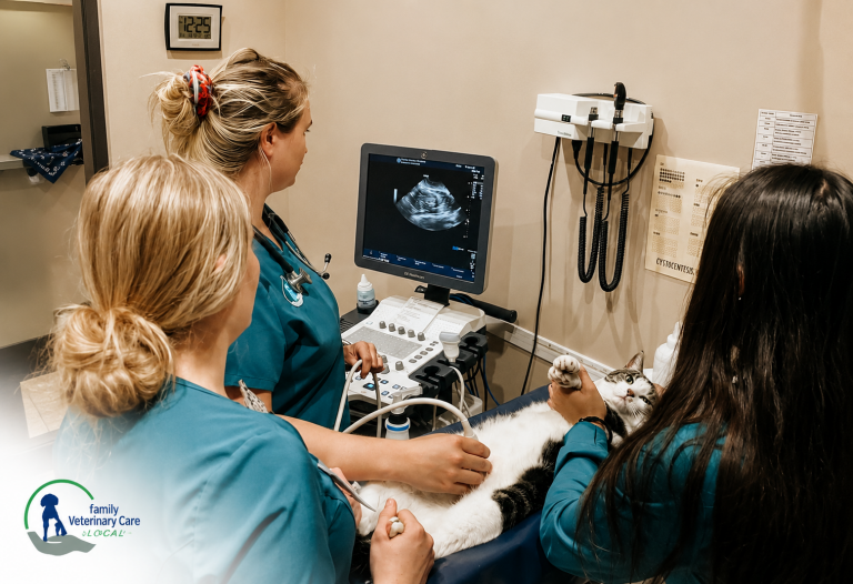
Pet Ultrasound Services: The Picture Inside the Picture
X-rays are excellent for dense structures like bone and for getting an overview of organ position and size. What they do not show as clearly is what is happening inside soft tissue organs. That is where pet ultrasound services become essential.
Ultrasound uses sound waves to generate real-time images of internal structures. It is completely non-invasive and does not involve radiation. For pets, it is especially useful for evaluating the liver, kidneys, spleen, bladder, and gastrointestinal tract in detail. It allows us to see whether a mass has defined borders or irregular edges, whether fluid is present in a body cavity, and whether organ texture and blood flow appear normal.
A study published in the Journal of Veterinary Internal Medicine found that abdominal ultrasound significantly increased diagnostic effectiveness for dogs and cats presenting with vague gastrointestinal signs, detecting conditions that would have been missed or delayed with physical examination alone.
Ultrasound is also used to guide procedures like fine needle aspirates, where a small sample of tissue or fluid is collected for analysis. Rather than guessing at location, the vet can watch in real time to ensure the needle is placed precisely where it needs to be. This makes sampling safer and more accurate.
Diagnostic Tests for Dogs and Cats: What Imaging Finds That Bloodwork Misses
Bloodwork and imaging are complementary, not interchangeable. Blood panels tell us about organ function, infection markers, blood cell counts, and metabolic values. What they cannot tell us is where the problem is physically located, how large it is, or whether it has affected surrounding structures.
A dog with elevated liver values on bloodwork needs imaging to understand why. Is there a mass? Is the liver enlarged uniformly, or is there a focal lesion? Is there fluid around the liver? These are structural questions that only imaging can answer, and they change the treatment plan entirely depending on the response.
Diagnostic tests for dogs and cats work best as a system. We use bloodwork to identify what is happening biochemically, imaging to locate and characterize the problem structurally, and then combine both to build a complete clinical picture. Relying on one without the other, especially in older pets or pets with vague symptoms, means accepting an incomplete diagnosis.
Early Disease Detection: Why the Timing of Imaging Matters
The value of imaging is not just in what it finds. It is in when it finds it.
Conditions like cancer, heart disease, kidney disease, and certain liver conditions produce few or no outward symptoms in their early stages. By the time a pet is visibly unwell, the disease has frequently progressed to a point where treatment is more aggressive, more expensive, and less likely to achieve a full recovery. Early imaging, whether at a wellness visit or at the first sign of something subtle, catches these conditions while they are still manageable.
An older dog who seems fine but is drinking more water than usual might have a splenic mass that shows up clearly on ultrasound before it ruptures. A cat who has lost a little weight might have a small intestinal thickening that points toward inflammatory bowel disease or early lymphoma. A dog with a slight cough might have early cardiac enlargement visible on a chest X-ray long before the cough becomes persistent or the heart failure advances.
In each of these cases, the imaging finding does not change what was already there. It changes what gets done about it, and when.
Veterinary Diagnostic Imaging at Family Veterinary Care of Oakdale
Family Veterinary Care of Oakdale offers digital radiography and ultrasound as part of our in-house diagnostic functions. This means we are not sending you to a referral center to wait for imaging that should be part of the same visit. When your pet needs a chest X-ray or an abdominal ultrasound, we do it here, interpret it here, and have a conversation with you about what it shows before you leave.
Our digital radiography system produces sharp images that allow us to evaluate bone and soft tissue detail clearly. For ultrasound, we use the real-time imaging to assess organ structure and guide any sampling that is needed. Images can be shared electronically with veterinary radiologists or specialists when a second read or a specialist opinion adds value to the case.
We use imaging as part of wellness care for senior pets, not just for sick visits. An older dog or cat who has not had imaging in a few years may have changes developing that bloodwork alone would not catch. Building imaging into senior care procedures is one of the most practical things we can do to find problems early rather than react to them late.
Pet owners from Oakdale, Riverbank, Escalon, Del Rio, Modesto, Manteca, Ripon, Turlock, and Stockton regularly bring their pets to us for diagnostic evaluations. If your pet needs imaging and you are looking for a clinic that handles it in-house with same-visit results, we are easy to reach and set up appointments without long lead times.
Conclusion
Pet diagnostic imaging is not a luxury add-on for complicated cases. It is one of the most direct tools available for finding disease early, making accurate diagnoses, and directing treatment decisions that actually fit what is happening inside your pet. The earlier a problem is identified, the more that can be done about it. If your dog or cat is due for a wellness visit, showing subtle symptoms you cannot quite explain, or has a condition that needs ongoing monitoring, call Family Veterinary Care of Oakdale at (209) 847-9077 or book online. Getting the full picture sooner rather than later is almost always the better call.
FAQs
Does my pet need to be sedated for X-rays or ultrasound?
Most pets do not require sedation for imaging. X-rays take only a few minutes, and many dogs and cats tolerate the positioning well. Sedation may be recommended for pets that are in significant pain, highly anxious, or need a position that is difficult to hold comfortably. We assess each pet individually and only use sedation when it genuinely improves the quality of the images and the experience for your pet.
How do I know if my pet needs imaging or just bloodwork?
Bloodwork and imaging answer different questions. Bloodwork tells us about organ function and systemic markers. Imaging shows us what is happening structurally. For many presentations, including unexplained weight loss, vomiting, lethargy, or a lump your vet can feel, both are needed to get a complete picture. Your vet will recommend based on what the exam findings show, but if there is any structural question involved, imaging provides information that bloodwork cannot.
Can Family Veterinary Care of Oakdale do imaging on the same day as the appointment?
Yes. We have digital radiography and ultrasound in-house, so imaging is performed and reviewed during the same visit whenever possible. You will leave knowing what the images displayed rather than waiting days for results from an outside facility.
Is pet ultrasound safe? Does it involve radiation?
Ultrasound uses sound waves, not radiation, and is completely safe for pets. It is non-invasive, painless, and does not have any known side effects. Digital X-rays do involve a small amount of radiation, but the exposure is minimal and considered safe for diagnostic purposes. We follow standard positioning and shielding procedures to keep exposure as low as possible.
My pet seems healthy. Is imaging still useful at a wellness visit?
For adult and senior pets, yes. Many conditions develop without obvious symptoms in their early stages. Including imaging in senior wellness care, particularly chest X-rays and abdominal ultrasound, allows us to catch changes before they become clinical problems. It is the same logic as routine blood panels: you are not checking because something is wrong; you are checking so you know before something becomes wrong.
Which cities near Oakdale do you serve for diagnostic imaging?
We see pets from Oakdale and the surrounding area, including Riverbank, Escalon, Del Rio, Modesto, Manteca, Ripon, Turlock, and Stockton. If you are looking for in-house veterinary diagnostic imaging without a long wait or a referral center trip, we are centrally located and take appointments for both new and existing patients.


Leave A Comment